Home » Without Label » Knee Muscle Anatomy Mri / MRI KNEE JOINT ANATOMY : The knee joint is a synovial joint which connects the femur thigh bone the longest bone in the body to the tibia shin bone.
Knee Muscle Anatomy Mri / MRI KNEE JOINT ANATOMY : The knee joint is a synovial joint which connects the femur thigh bone the longest bone in the body to the tibia shin bone.
Knee Muscle Anatomy Mri / MRI KNEE JOINT ANATOMY : The knee joint is a synovial joint which connects the femur thigh bone the longest bone in the body to the tibia shin bone.. Free access interactive and dynamic anatomical atlas. This mri knee sagittal cross sectional anatomy tool is absolutely free to use. Three conventional mri planes that are utilized to evaluate the knee include sagittal (oblique), coronal, and transaxial planes. The muscles that affect the knee's movement run along the thigh and calf. These muscles work in groups to flex extend and stabilize the knee joint.
Atlas of knee mri anatomy. Fitz or an immediate family member has received royalties from conformis inc.; Anatomy_of_knee_mri 3/3 anatomy of knee mri anatomy of knee mri recognizing the quirk ways to acquire this ebook anatomy of knee mri is additionally useful. The femur, tibia and patella.the arrangement of the bones in the knee joint, along with its many ligaments, provide it with the arthrokinematics that allows for great stability, combined with great mobility.being arguably the most stressed and exposed joint of the body, the knee joint is predisposed to various. Anatomy basic knee mri checklist.
Knee Muscle Anatomy Mri - Atlas of Knee MRI Anatomy - W ... from image.slidesharecdn.com Mri knee anatomy scroll using the mouse wheel or the arrows. These muscles work in groups to flex, extend and stabilize the knee joint. T2w axial fat sat 1. Magnetic resonance imaging (mri scan): Mri wrist anatomy scroll using the mouse wheel or the arrows. Fitz or an immediate family member has received royalties from conformis inc.; Thigh muscles also protect neurovascular structures as they go through the proximal hip joint to the knee and lower leg (3). In this presentation mri anatomy biceps femoris muscle.
There is a flat area of tendon originating from the knee.
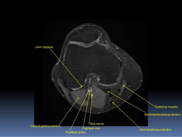
Weak adductor muscles may cause knee instability and adductor strain (2). Richolt j.a., jakab m., kikinis r. Three conventional mri planes that are utilized to evaluate the knee include sagittal (oblique), coronal, and transaxial planes. In approximately 2% of the population, the anterior tibial artery branches along the keywords: The knee joint is a modified hinge joint between the femur, tibia, and patella. Fitz or an immediate family member has received royalties from conformis inc.; Related posts of knee muscle anatomy mri anatomy muscle system. Atlas of knee mri anatomy. Assoc prof craig hacking and dr shu su et al. This article is based on a presentation given by david rubin and adapted for the radiology assistant by robin smithuis. Knee muscle anatomy axial mri : Anatomy of the knee bones around the knee. Injuries such as anterior cruciate ligament, meniscus and rotator cuff tears are all easily diagnosed when there is a firm understanding and knowledge of human anatomy.
Magnetic resonance imaging (mri scan): The muscles that affect the knee's movement run along the thigh and calf. Mri wrist anatomy scroll using the mouse wheel or the arrows. Atlas of knee mri anatomy. Mri knee anatomy scroll using the mouse wheel or the arrows.
Pin on Mri from i.pinimg.com The images may also help physicians to distinguish normal, healthy tissues from dead tissues(2). Weak adductor muscles may cause knee instability and adductor strain (2). These muscles work in groups to flex, extend and stabilize the knee joint. This mri knee sagittal cross sectional anatomy tool is absolutely free to use. In this presentation mri anatomy biceps femoris muscle. They are attached to the femur (thighbone), tibia (shinbone), and fibula (calf bone) by fibrous tissues called ligaments. Thigh muscles also protect neurovascular structures as they go through the proximal hip joint to the knee and lower leg (3). The patellofemoral articulation, consisting of the patella, or kneecap, and the patellar groove on the front of the femur through which it slides;
The images may also help physicians to distinguish normal, healthy tissues from dead tissues(2).
The muscles that affect the knee's movement run along the thigh and calf. The patellofemoral articulation, consisting of the patella, or kneecap, and the patellar groove on the front of the femur through which it slides; Atlas of anatomy in medical imagery. Mri wrist anatomy scroll using the mouse wheel or the arrows. When a muscle has different orientations of the tendons it means that there are different patterns of edema possible depending on the tendon injured. Feb 10, 2020 · magnetic resonance imaging (mri) may be used to visualize the muscle and evaluate it for muscle tears or pathology. Injuries such as anterior cruciate ligament, meniscus and rotator cuff tears are all easily diagnosed when there is a firm understanding and knowledge of human anatomy. Medical images from an mri allow medical professionals to distinguish body tissues, including the meniscus (shock absorbers in the knee), cartilage, tendons, and ligaments. Stanford msk mri atlas, radlex Superiorly, it extends to the level of the crossing of the biceps femoris tendon, and remains superficial to fcl in this location.10 Anatomy basic knee mri checklist. Louis, usa and the rijnland hospital in leiderdorp, the netherlands. Use the mouse scroll wheel to move the images up and down alternatively use the tiny arrows (>>) on both side of the image to move the images.>>) on both side of the image to move the images.
This article is based on a presentation given by david rubin and adapted for the radiology assistant by robin smithuis. Medical images from an mri allow medical professionals to distinguish body tissues, including the meniscus (shock absorbers in the knee), cartilage, tendons, and ligaments. Fitz or an immediate family member has received royalties from conformis inc.; Anatomy arthrogram anatomy basic shoulder mri. Knee mri anatomy of the knee anterior cruciate ligament pet ct journal prompts biceps study health fitness.
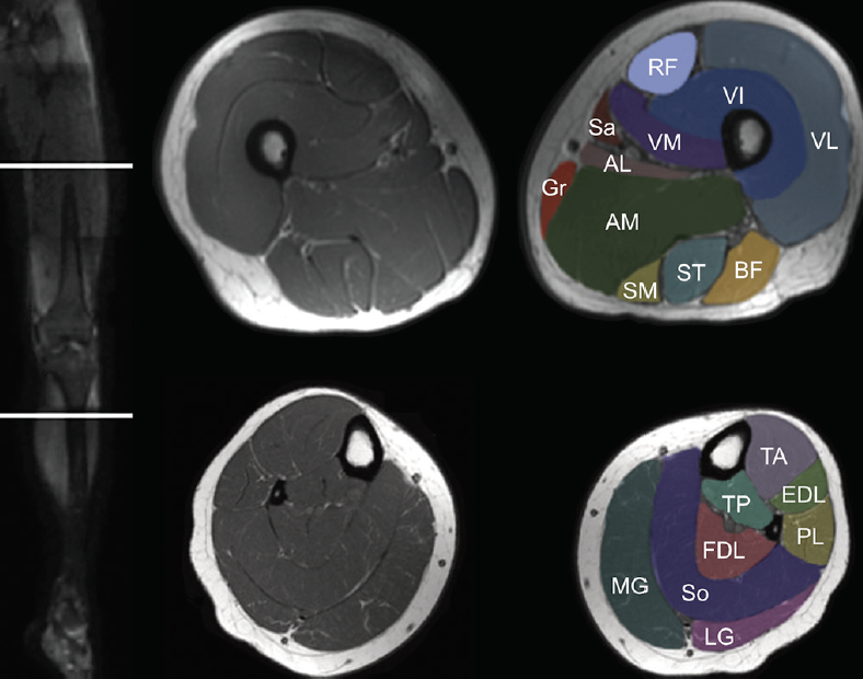
Knee Muscle Anatomy Mri / Mri Knee Joint Anatomy - Find ... from core4.bmctoday.net Anatomy muscle system 12 photos of the anatomy muscle system anatomy and physiology muscular system exam, anatomy and physiology muscular system labeling quiz, anatomy and physiology muscular system pdf, anatomy and physiology muscular system review, human anatomy muscular system quizzes, human muscles, anatomy and physiology. Mri knee anatomy scroll using the mouse wheel or the arrows. Knee mri anatomy of the knee fascia lata windows server muscles surface education muscle onderwijs. Abnormal anatomy with normal signal, i.e. Atlas of knee mri anatomy. This mri hip joint axial cross sectional anatomy tool is absolutely free to use. Anatomy basic knee mri checklist. An mri of the knee of a healthy subject was performed in the 3 planes of space (coronal, axial, sagittal) commonly used in osteoarticular imaging, with two weightings most commonly used to explore the musculoskeletal pathology of the knee:
The knee joint is the junction of the thigh.
Knee mri anatomy of the knee fascia lata windows server muscles surface education muscle onderwijs. Assoc prof craig hacking and dr shu su et al. Magnetic resonance imaging (mri scan): In approximately 2% of the population, the anterior tibial artery branches along the keywords: Magnetic resonance imaging (mri scan): Atlas of anatomy in medical imagery. Knee mri anatomy of the knee anterior cruciate ligament pet ct journal prompts biceps study health fitness. Knee mri, popliteal vessels, vascular. This mri knee sagittal cross sectional anatomy tool is absolutely free to use. Thigh muscles also protect neurovascular structures as they go through the proximal hip joint to the knee and lower leg (3). Stanford msk mri atlas, radlex An mri of the knee of a healthy subject was performed in the 3 planes of space (coronal, axial, sagittal) commonly used in osteoarticular imaging, with two weightings most commonly used to explore the musculoskeletal pathology of the knee: Anatomy of the knee bones around the knee.